Radigraph
Panoramic X-rays
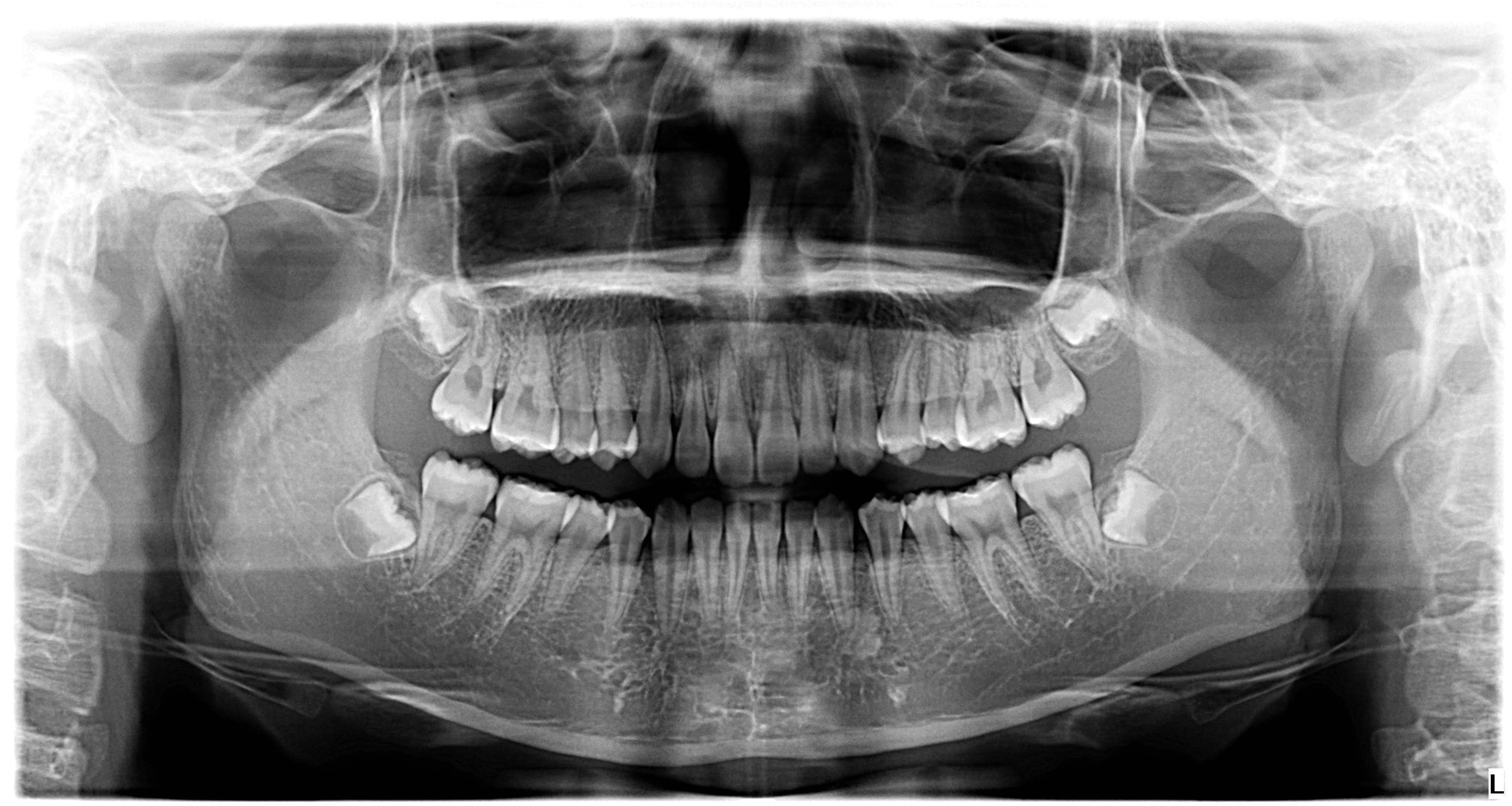
Panoramic X-rays (also known as Panorex® or orthopantomograms) are wraparound photographs of the face and teeth. They offer a view that would otherwise be invisible to the naked eye. X-rays in general, expose hidden structures, such as wisdom teeth, reveal preliminary signs of cavities, and also show fractures and bone loss.
Panoramic X-rays are extraoral and simple to perform. Usually, dental X-rays involve the film being placed inside the mouth, but panoramic film is hidden inside a mechanism that rotates around the outside of the head.
Unlike bitewing X-rays that need to be taken every few years, panoramic X-rays are generally only taken on an as-needed basis. A panoramic X-ray is not conducted to give a detailed view of each tooth, but rather to provide a better view of the sinus areas, nasal areas and mandibular nerve. Panoramic X-rays are preferable to bitewing X-rays when a patient is in extreme pain, and when a sinus problem is suspected to have caused dental problems.
Panoramic X-rays are extremely versatile in dentistry, and are used to:
- Assess patients with an extreme gag reflex.
- Evaluate the progression of TMJ.
- Expose cysts and abnormalities.
- Expose impacted teeth.
- Expose jawbone fractures.
- Plan treatment (full and partial dentures, braces and implants).
- Reveal gum disease and cavities.
How are panoramic X-rays taken?
The panoramic X-ray provides the dentist with an ear-to-ear two-dimensional view of both the upper and lower jaw. The most common uses for panoramic X-rays are to reveal the positioning of wisdom teeth and to check whether dental implants will affect the mandibular nerve (the nerve extending toward the lower lip).
The Panorex equipment consists of a rotating arm that holds the X-ray generator, and a moving film attachment that holds the pictures. The head is positioned between these two devices. The X-ray generator moves around the head taking pictures as orthogonally as possible. The positioning of the head and body is what determines how sharp, clear and useful the X-rays will be to the dentist. The pictures are magnified by as much as 30% to ensure that even the minutest detail will be noted.
Panoramic X-rays are an important diagnostic tool and are also valuable for planning future treatment. They are safer than other types of X-rays because less radiation enters the body.
If you have questions or concerns about panoramic X-rays, please contact our practice.
Cephalometric X-rays
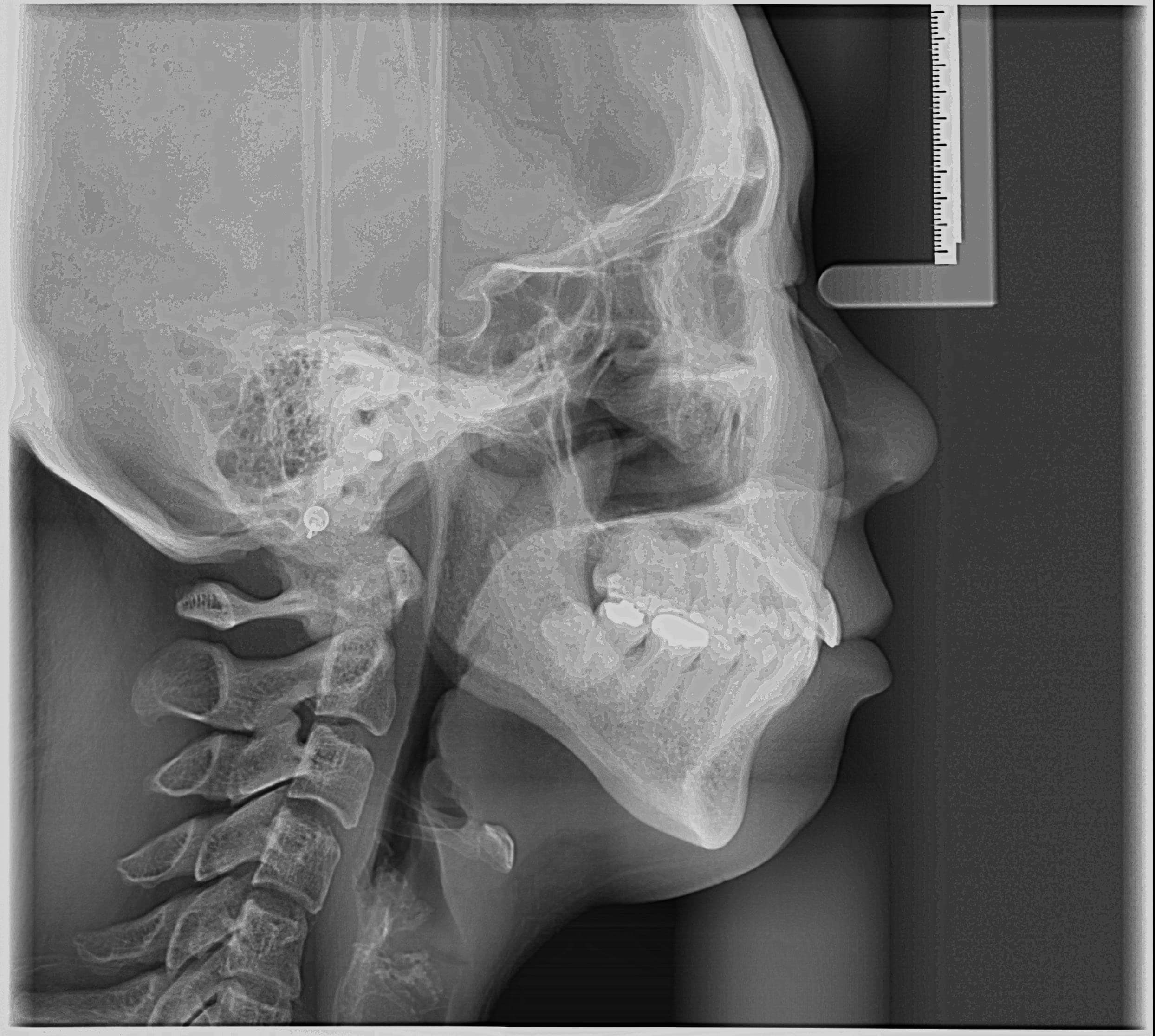
The cephalometric X-ray is a unique tool that enables the dentist to capture a complete radiographic image of the side of the face. X-rays in general offer the dentist a way to view the teeth, jawbone, and soft tissues beyond what can be seen with the naked eye. Cephalometric X-rays are extraoral, meaning that no plates or film are inserted inside the mouth. Cephalometric and panoramic X-rays display the nasal and sinus passages, which are missed by intraoral bitewing X-rays.
Cephalometric X-rays are usually taken with a panoramic X-ray machine. The adapted machine will have a special cephalometric film holder mounted on a mechanical arm. An X-ray image receptor is exposed to ionizing radiation in order to provide the dentist with pictures of the entire oral structure. The advantage of both cephalometric and panoramic X-rays is that the body is exposed to less radiation.
Cephalometric X-rays are not as common as “full sets” or bitewing X-rays, but they serve several important functions:
- Provide views of the side profile of the face.
- Provide views of the jaw in relation to the cheekbone.
- Provide information about “bad bites” or malocclusions.
- Allow measurement of the teeth.
- Identify fractures and other injuries to the teeth and jawbone.
- Assists in orthodontic planning.
How are cephalometric X-rays taken?
Cephalometric X-rays are completely painless. The head is placed between the mechanical rotating arm and the film holder, which is placed on another arm. The arm rotates around the head capturing images of the face, mouth, and teeth. The clarity and sharpness of these images will depend on the positioning of the body. The images are usually magnified up to 30%, so any signs of decay, disease, or injury can be seen and treated.
After capturing cephalometric X-rays, the dentist will be able to see a complete side profile of the head. This can assist in orthodontic planning and allow an immediate evaluation of how braces might impact the facial profile and teeth. Another common use for this type of X-ray is to determine specific measurements prior to the creation and placement of dental implants.
If you have any questions or concerns about cephalometric X-rays, please ask your dentist.